
Starfish have a complete digestive system with a mouth at the center of their underside (the "oral" side) and an anus on their upper surface (the "aboral" side). The figure below (from Brusca and Brusca) is a cut-away view of the digestive system and associated structures. The top of the starfish is up; one ray extends off to the right.
Food can be brought into the stomach through the mouth or, in many species, the cardiac stomach can be extended out through the mouth to digest food outside the body. Suspension-feeding starfish use their tube feet to pass food to the mouth. The cardiac stomach is connected to a pyloric stomach (located above it), which in turn is connected to both the anus and to the pyloric ducts and pyloric cecum which extend out into each arm (see the figure below).
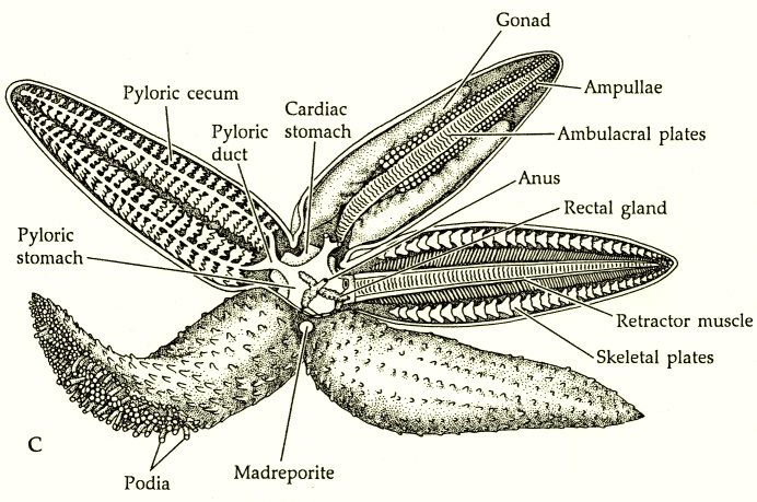
The pyloric ceca (or digestive glands) and the cardiac stomach produce digestive enzymes. Digested material is absorbed through the pyloric ceca for transport to the rest of the body. Since the pyloric ceca extend the length of each arm, the need for an advanced circulatory system is reduced.
Circulation occurs in three places: the perivisceral coelom (basically, the space inside the body but outside the various organs), the water vascular system (of which the tube feet are the most obvious part), and the hemal system (which actually looks something like a circulatory system). The hemal system is shown below. There are hemal channels forming rings around the central part of the body around the mouth (the oral hemal ring), closer to the upper surface (the aboral hemal ring), and a third ring around the digestive system (the gastric hemal ring). These are connected by the axial sinus. There are also radial hemal channels running down the rays next to the gonads (which are also located in the rays). A dorsal sac attached to the axial sinus pulsates, sort of like a very inefficient heart (inefficient because it lacks a one-way valve system). The hemal system seems mostly organized to distribute nutrients from the digestive tract.

The water vascular system uses cilia and the constant contraction of ampulla (to extend and retract the tube feet) also helps keep things moving. There is an ionic imbalance that causes water to flow into the water vascular system through the madreporite, and then the Tiedemann's bodies divert some of it into the perivisceral coelom. Circulation in the perivisceral coelom is mostly by ciliary beating.
 |
Oxygen enters mainly by diffusion into the tube feet (and thus into the water vascular system) or the papulae, which are little sacs all over the upper surface of the body. The figure to the left is a close-up of the aboral surface of a sunflower star. The pincer-like structures, called pedicellariae, are probably used for defense and in some species can capture small food particles. All of the smaller sac-like structures are papulae. You can see these structures on starfish with your eyes, but it's easier with a magnifying glass or dissecting microscope. |
Back to my homepage |
Last updated May 10, 2000.
